Mammography

Mammography is the process of using low-energy X-rays to examine the human breast for diagnosis and screening. The goal of mammography is the early detection of breast cancer, typically through detection of characteristic masses or microcalcifications.
As with all X-rays, mammograms use doses of ionizing radiation to create images. These images are then analyzed for abnormal findings. It is usual to employ lower-energy X-rays than those used for radiography of bones. Mammography may be 2D or 3D (tomosynthesis). Ultrasound is typically used for further evaluation of masses found on
mammography or palpable masses that may or may not be seen on
mammograms.
Type of Mammography;
-
Digital Mammography 2D
-
3D Mammography (Tomosynthesis)
Source: Wikipedia
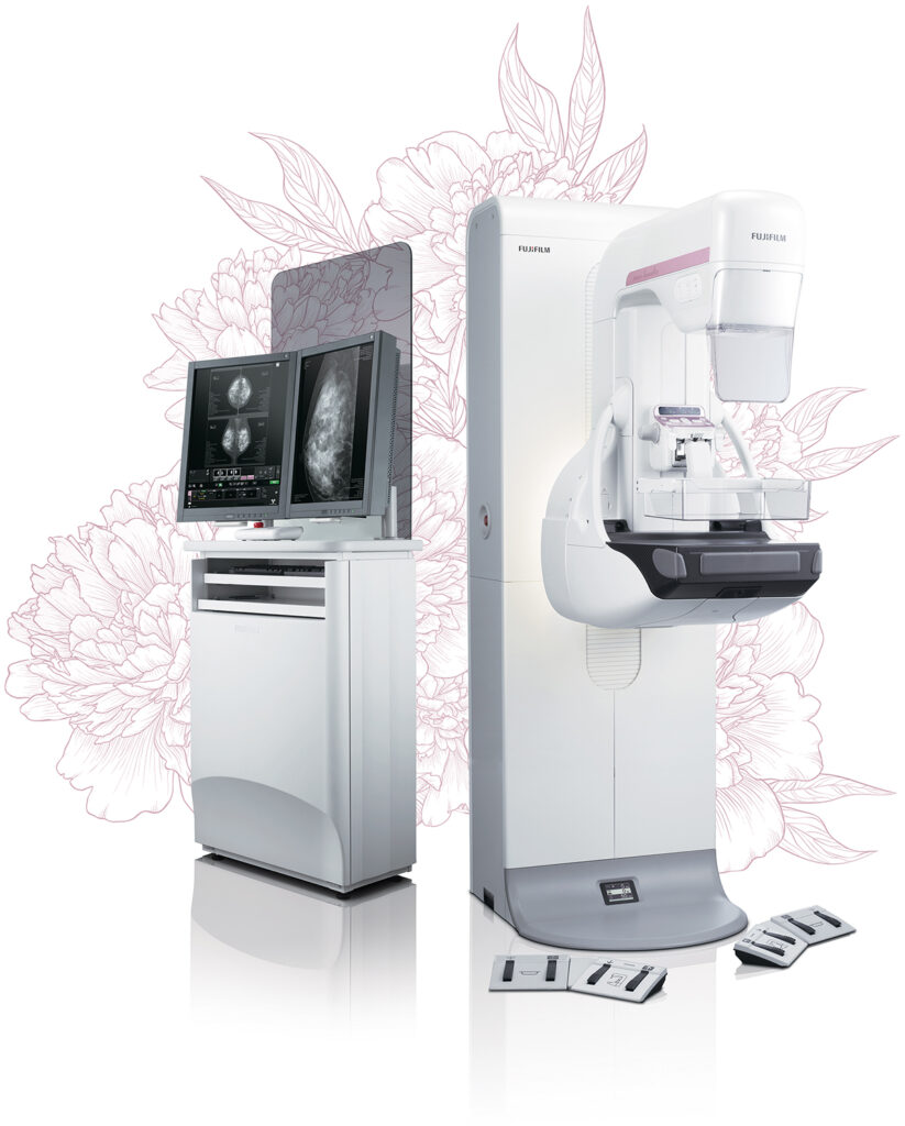
Fujifilm AMULET Innovality Mammography
Unique detector for fast, low dose examinations
AMULET Innovality employs a direct-conversion flat panel detector made of Amorphous Selenium (a-Se) which exhibits excellent conversion efficiency in the mammographic X-ray spectrum. The HCP (Hexagonal Close Pattern) detector efficiently collects electrical signals converted from X-rays to realize both high resolution and low noise. This unique design makes it possible to realize a higher DQE (Detective Quantum Efficiency) than with the square pixel array of conventional TFT panels. With the information collected by the HCP detector, AMULET Innovality creates high definition images with a pixel size of 50 μm; the finest available with a direct-conversion detector.
This low-noise and high-speed switching technology allows tomosynthesis exposures with a low X-ray dosage and short acquisition time to be performed. Fast image display is also possible, realizing a smooth mammography workflow from exposure to image display.
Tomosynthesis: making it possible to observe the internal structure of the breast
In breast tomosynthesis, the X-ray tube moves through an arc while acquiring a series of low-dose X-ray images.
The images taken from different angles are reconstructed into a range of Tomosynthesis slices where the structure of interest is always in focus.
The reconstructed tomographic images make it easier to identify lesions which might be difficult to visualize in routine mammography because of the presence of overlapping breast structures.
Automatic compression reduction control (Comfort Comp)
This function will reduce the compression pressure within a range (within + 3 mm) in which the thickness of the breast does not change after normal breast compression is completed for the purpose of alleviating the patient’s pain.
AMULET Harmony incorporates a range of mammography solutions specifically designed to maintain a harmonious examination environment and foster an atmosphere of trust between mammographers and their patients.
Courtesy: Fujifilm India Private Limited

