Ultrasound


Ultrasound is composed of sound waves with frequencies greater than 20,000 Hz, which is by approximation the upper threshold of human hearing. Ultrasonic images, also known as sonograms, are created by sending pulses of ultrasound into tissue using a probe. The ultrasound pulses echo off tissues with different reflection properties and are returned to the probe which records and displays them as an image.
A general-purpose ultrasonic transducer may be used for most imaging purposes but some situations may require the use of a specialized transducer. Most ultrasound examination is done using a transducer on the surface of the body, but improved visualization is often possible if a transducer can be placed inside the body. For this purpose, special-use transducers, including transvaginal, endorectal, and transesophageal transducers are commonly employed. At the extreme, very small transducers can be mounted on small diameter catheters and placed within blood vessels to image the walls and disease of those vessels.
Compared to other medical imaging modalities, ultrasound has several advantages. It provides images in real-time, is portable, and can consequently be brought to the bedside. It is substantially lower in cost than other imaging strategies. Drawbacks include various limits on its field of view, the need for patient cooperation, dependence on patient physique, difficulty imaging structures obscured by bone, air or gases, and the necessity of a skilled operator, usually with professional training.
Uses:
Neonatology
Ophthalmology (eyes)
Source: Wikipedia
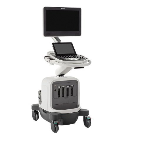
Philips Affiniti 30, 50 and 70 Machines
Flow Viewer
Anatomical Intelligence for Breast
Next Gen AutoSCAN
Philips Next Gen AutoSCAN improves image uniformity, adaptively adjusting image brightness at every pixel and reducing the need for user adjustment while also improving transducer plunkability. Next Gen AutoSCAN reduces button pushes by up to 54% with pixel-by-pixel real-time optimization.
MFI
TrueVue advanced 3D display
Fusion and Navigation
PureWave & xMATRIX transducer technology
Courtesy: Philips India Limited
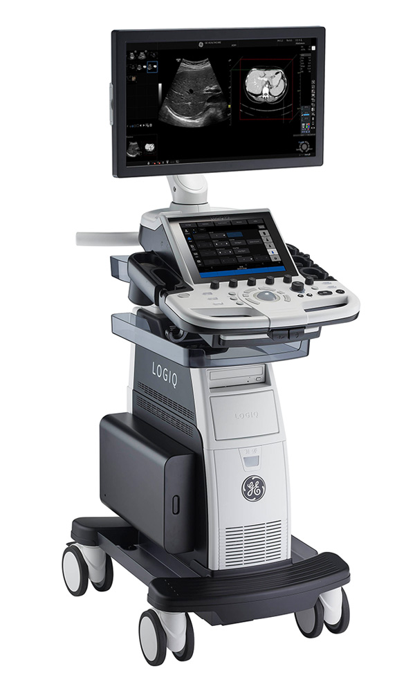
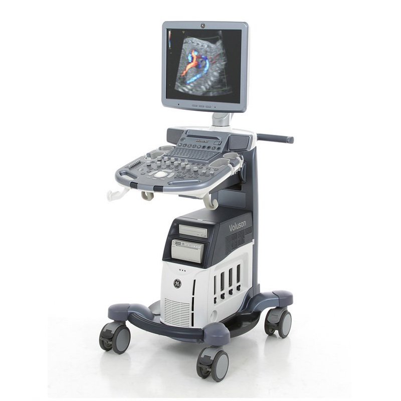
Wipro GE P9 R3 and Voluson S8 Machines
Expanded patient-centric diagnostic capabilities for flexibility
Multi-purpose capabilities, including liver, cardiac, OB/GYN, vascular, breast, thyroid, musculoskeletal, urologic, and pediatric studies.
Superb image quality with XDclear probes: Powerful high fidelity and broad bandwidth produce high resolution images whether scanning superficial or deep targets.
Advanced imaging and visualization tools, including:
• 2D Shear Wave Elastography
• Ultrasound-Guided Attenuation Parameter (UGAP)
• CEUS
• B-Flow and B-Flow Color
• 3D/4D with SonoRenderlive
• Stress Echo
Personalized workflow tools and automation for efficiency
Intuitive user interface – With customizable keys, joystick, and touch control, the system operates intuitively, enabling you to complete exams with fewer keystrokes.
Personalized set-up tools – With Start Assistant and My Preset, users can customize their own workflow preferences and use case presets, and then launch these settings in seconds.
Automated scanning tools – Continuous Tissue Optimization (CTO), Auto IMT, AutoEF, Measure Assistant, Compare Assistant, and Scan Assistant help reduce exam time and increase user efficiency.
HD-Flow™
Bi-directional Doppler feature that reduces overwriting and increases sensitivity over conventional color Doppler.
SonoVCAD™heart
Automatically
extracts and displays the standard recommended cardiac views from a single STIC (Spatio-Temporal Imaging Correlation) volume acquisition.
Tomographic Ultrasound Imaging (TUI)
Helps simplify analysis and documentation of dynamic studies by simultaneously displaying views of multiple parallel slices of a volume data set.
HDlive™ Technologies
Utilize a combination of advanced skin illuminating and shadowing techniques to display images with unprecedented depth and clarity.
Volume Contrast Imaging (VCI)
Adjusts slice thickness on 3D or 4D images to help enhance contrast resolution using bone and tissue rendering techniques.
SonoAVC™follicle & SonoAVCantral
SonoAVC™follicle & SonoAVCantral automatically calculates the number, dimensions, and volume of hypoechoic structures for follicle monitoring or antral follicle count.
Scan Assistant
This
flexible, customizable exam protocol helps reduce time required to
conduct and document results by guiding you through an exam more
efficiently, aiding in annotation, measurement, and reporting.

